Cytoskeletal Changes in T Cell Activation
Cytoskeletal Changes in T Cell Activation
Submitted by Sudha Kumari (Irvine Laboratory, KI at MIT) and Zsolt Lazar (Cold Spring Harbor Laboratories)
MIT Department of Biological Engineering, MIT Department of Materials Science and Engineering, Koch Institute at MIT
The T cells are round cells that carry out immunosurveillance, while passing through various organs of immune system. A unique property of immune cells is to be able to change their morphology remarkably in a matter of seconds as soon as they sense pathogen or foreign proteins. Encounter with antigen is quite like a raw egg hitting a hot pan, resulting into omelet within a matter of seconds. I am interested in studying the early events of T cell activation.
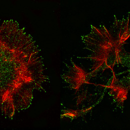
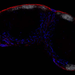
The image shows T cells in two stages of sensing and activation. The image is shown from bottom perspective such that the contact area is in focus. Left cell shows beginning of the attachment of T cell to a surface coated with foreign peptide (red, <30 sec attachment), the cell on the right shows later stage of stable attachment (<1 min of attachment), with large amount of foreign protein gathered underneath. Green pseudocolor shows T cell cytoskeletal framework. T cells possess quite unique cytoskeletal properties.

