An Inside View of Neural Tube Defects
An Inside View of Neural Tube Defects
Submitted by Hao Yin of the Langer and Anderson Laboratories at the Koch Institute for Integrative Cancer Research at MIT
Koch Institute at MIT, MIT Department of Chemical Engineering, Institute of Medical Engineering and Science
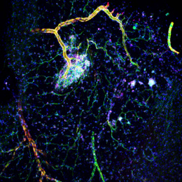
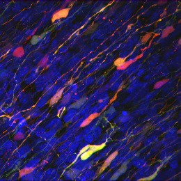
Neural tube defects (NTDs) are one of the most common congenital defects occurring in 1 in 1000 live births in humans. NTDs result from incomplete closure of the neural tube during early development, E8.5-9.5 in the mouse and weeks 3-4 in human. We are interested in learning the development process of neural tube closure. This is a mouse embryonic neural tube (E10.5), which will develop into brain during embryogenesis. By staining the neural tube with different markers, it helps us to understand the composition of neural tube and study the cellular behavior of different cell population.
Red (E-cadherin): neural epithelium. Yellow (Arl13b): cilia. Pink (p-histone3): proliferating cell at ventricular zone. white (islet1/2): motor neuron. Green (Tuj1): differentiated cells
The identification of genes required for the regulation of neural tube development in the mouse provides invaluable information including predicting candidate genes that contribute to human NTDs as well as determining how these genes function in the complex morphogenic process of neural tube closure.


