Dendritic Cell Motion Within a Tumor 2
Dendritic Cell Motion Within a Tumor 2
Tim Fessenden
MIT Department of Biology, Koch Institute at MIT
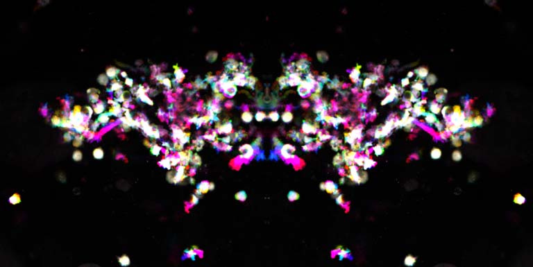
The image depicts very specialized of immune cells, called dendritic cells, as they move inside the tumor of a living mouse. These images were taken as a time series over 30 minutes. Time has been encoded as colors in an overlay. Immobile parts of the dendritic cells are white while cell movements are shown as different colors, depending on their exact timing.
Dendritic cells can direct the immune system to eradicate a tumor, but precisely how they exert their effects and how they go wrong is not well understood. My work attempts to understand dendritic cell biology through microscopy, by recording dendritic cell number and movements inside the same tumor on consecutive days. As those tumors are either rejected or able to overcome the mouse immune response and continue growing, I record what dendritic cells are doing inside the tumor to connect their behavior with the final outcome. In these cases, the two image series I used showed active dendritic cells, and the tumors of these mice were eventually cleared completely by the immune system.
