Remodel Organism: Tissue Origami in a Fruit Fly Embryo
Remodel Organism: Tissue Origami in a Fruit Fly Embryo
Collections: Image Award Winners
2023 Award Winner
Mary Ann Collins, Adam Martin
MIT Department of Biology
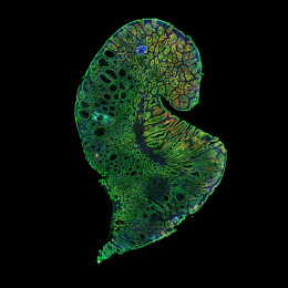
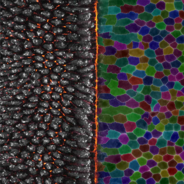
As an organism transforms from a simple collection of cells into a complex system of life, the developing tissue undergoes extensive remodeling. The Martin Lab uses fruit fly embryos to study the impact of mechanical forces on cell behavior.
On the left, nuclei in gray are linked by new cell junctures, marked in orange. The view on the right maps cell boundaries with randomly assigned colors to track them as they evolve. At center, a newly-formed structure fold pulls the two sides inward. Collectively, these perspectives paint a dynamic picture of a cellular community working together to shape the organism.
Video
Mary Ann Collins presents on her image.
