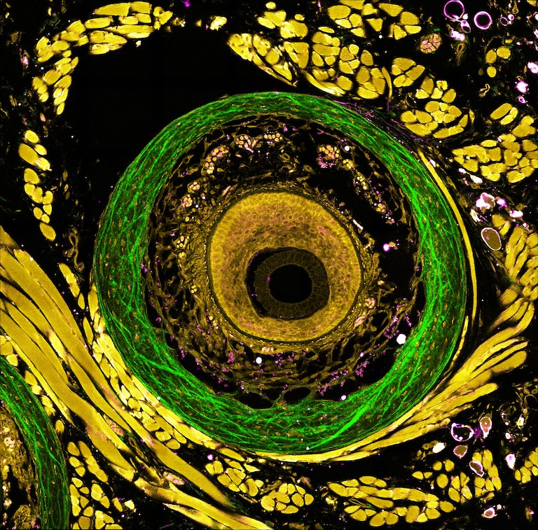
The Tissue Microenvironment of Unprocessed Mouse Whisker Pads
The Tissue Microenvironment of Unprocessed Mouse Whisker Pads
Kunzan Liu, Tong Qiu, Jiashu Han, Eva Lendaro, Manuel Levy, Ellen Kan, Keith Isaacson, Fan Wang, Linda Griffith, Sixian You
Koch Institute at MIT, Research Laboratory of Electronics
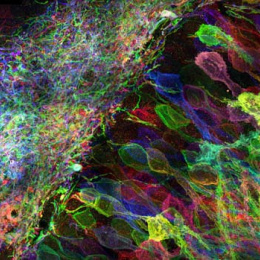
The overarching objective of these series of images (2 of 3) is to extend the boundaries of label-free optical microscopy, enabling label-free imaging at 10 Hz (20-times faster), probing depths of up to 2 mm (10-fold deeper), and offering more than three distinct colors for precise molecular and structural identification. With the developed imaging techniques, it has helped enhance the understanding of intricate diseases, therapeutic impacts, and biological processes with minimal disturbance to biological specimens.
For example, studying chronic neuropathic pain is crucial for alleviating patient suffering and improving the overall quality of life for affected individuals. By studying whisker pads, the hope is to uncover the chronic neuropathic pain related to the whisker sensory pathway. Through label-free multicolor profiling, the capability to discern various cell types and quantify their redox states based on their distinct colors and morphology will be obtained. This encompasses the analysis of stromal cells, adipocytes, muscle cells, immune cells, trabecula, and more, allowing us to infer changes occurring both before and after the injury.